Particle Research using AFM at Virginia Tech’s Nano Earth Lab
- 21 Aug 2019
- Volume 16
- NanoScientific Magazine, Spring 2019

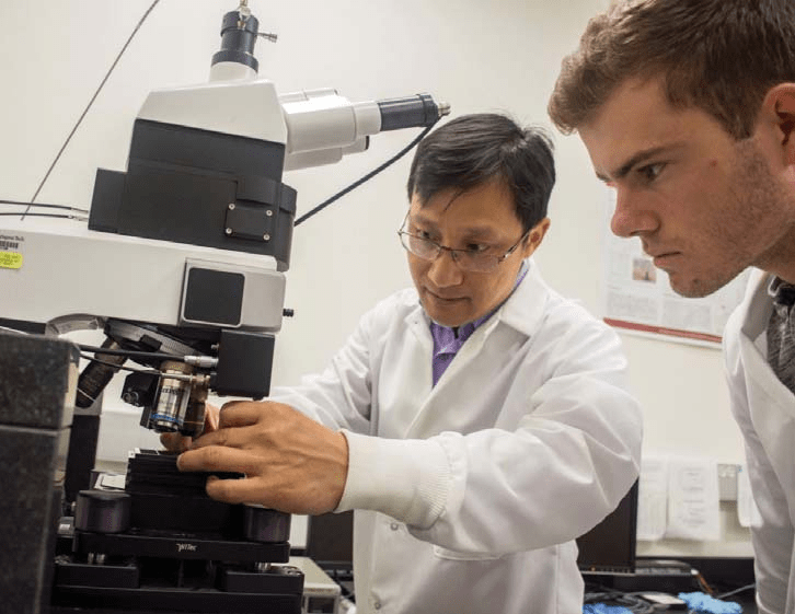
Pictured above, Dr. Weinan Leng, who researches Nanoparticle-based sensors for environmental applications and Laser-based techniques (Raman and surface enhanced Raman scattering) for nanoparticle tracking.
AT VIRGINIA TECH, ONE OF THE MEMBERS OF CEINT THE THE VIRGINIA TECH CENTER FOR SUSTAINABLE NANOTECHNOLOGY (VTSUN), IS FOR STUDENTS AND POSTDOCS WHO ARE RESEARCHING ENVIRONMENTAL NANOSCIENCE AND TECHNOLOGY FROM DIFFERENT PERSPECTIVES AND IS EQUIPPED WITH A RANGE OF STATE-OF-THE ART NANOMETROLODGY TOOLS INCLUDING ATOMIC FORCE MICROSCOPE, TEM AND SEM.
AN INTERVIEW WITH DR. WEINAN LENG, LABORATORY DIRECTOR OF VIRGINIA TECH NATIONAL CENTER FOR EARTH AND ENVIRONMENTAL NANOTECHNOLOGY INFRASTRUCTURE (NANOEARTH)
How do you use Atomic Force Microscopes in your research?
In most of cases, I utilize AFM for topographic measurements, such as particle size, surface roughness.
What features of Atomic Force Microscope are most useful and why?
The most useful feature of AFM is to measure incredible small particles with a high degree of accuracy.
Can you show an example of silver nano particle imaging and explain why these images were taken and what they tell us?
Figure 1. AFM images of uncoated and coated AgNPs measured after 1 and 14-days dissolution. The left panel is the AFM image for the original AgNPs without coating and dissolution; on the right side (labeled a-h) are the AFM images for uncoated and coated AgNPs after 1 and 14 days. (Liu, C., Leng, W., Vikesland, P. J., Controlled Evaluation of the Impacts of Surface Coating on Silver Nanoparticle Dissolution Rates, ES&T, 2018, 52(5),2726-2734.)
This is an example of how to utilize a highly sensitive AFM to examine engineered nanomaterial dissolution, which is one of NSF projects that has been done in the group of Prof. Peter Vikesland at Virginia Tech. The AFM images in Figure 1 illustrate how surface coatings affect silver nanoparticle dissolution rates. In this research, arrays of surface adhered silver nanoparticles were produced via nanosphere lithography, and AFM provides a convenient technique to measure changes in particle height which is related to the kinetics of AgNP dissolution.

Figure 1

Can you show an example of gold nano particle imaging and explain why the images were taken and what they tell us?
In Figure 2, on the next page, AFM images of 5nm gold nano particle (Au NP) standard (A) and 1 nm gold hydrogel sample (B).
This is one of the most attractive applications of AFM which provides a quick survey for very small particles (less than 5 nm) in a low concentration that are not created as an artifact of the electron beam in an electron microscope. The standard image shows how accuracy of height measurements which can reflect particle size.
How do you think the imaging of NanoParticles can help us with the environmental issues we are facing?
Following the example in question 3, once a well-controlled nano structure was designed and modelled for the engineered nano materials, which can be avoid the severe limitations of many alternative methodologies, such as the aggregation using realistic nanoparticle solution.
Findings based on AFM imaging will aid eff orts to engineer environmentally benign nanomaterials and to predict their environmental impacts.
What do you like best about working in the NanoEarth group at VaTech?
I love being an instrument specialist, and being asked for my participation in a lot of cool projects of NanoEarth users. Every technical service, data report and critical observation you give will have your fingerprint on it in a good way. It gives me great job satisfaction that significantly outweighs the stress, the time and the hard work.

Dr. Weinan Leng is a Laboratory Director of Virginia Tech National Center for Earth and Environmental Nanotechnology Infrastructure (NanoEarth), operating Raman-AFM microscope, X-ray photonelectron spectroscope, multimode AFM, BET, UV-vis-NIR spectrometer and DLS instrument. He received his PhD from Southeast University of China in Biomedical Engineering for his work on the fabrication and characterization of second-order nonlinear optical polymers and their application in electro-optical devices.
He moved to the United States as a post-doctoral researcher in the group of Prof. Anne Myers Kelly (University of California, Merced) working on the development of laser optical spectroscopes. From 2009 to 2018, Dr Leng was a Research Scientist at Virginia Tech, where he implements his research lines originated in Environmental Nanotechnology.
Since September 2018, Dr Leng is a senior Research Associate at the Institute for Critical Technology and Applied Science of Virginia Tech.