Cellulose NanofibrilsTopography & Young’s Modulus Imaging Using Park Atomic Force Microscope
- 16 Jun 2016
- Volume 7
- NanoScientific Magazine, Summer 2016
Byong Kim, Gerald Pascual, Phani Kondapani and Keibock Lee
Technical Marketing, Park Systems, Inc., Santa Clara, USA,
Abstract
Carbon nanotubes (CNTs) are a popular sample for various nanotechnology studies. However, they are very difficult to synthesize en masse. As a result, researchers have started looking at alternatives with similar properties. One of the candidates to replace CNTs in some nanocomposite materials is cellulose nanowhiskers (CNWs), structures that can be readily produced from plant sources. To determine the feasibility of substituting CNWs for CNTs, it is important to verify the two materials have similar morphologies and nanomechanical properties. One must first establish a baseline for comparison by accurately characterizing CNW properties at nanoscale. To this end, three CNW samples were examined using an atomic force microscope (AFM) with non-contact AFM mode and an AFM-based fast nanomechanical mode. The target properties for evaluation were the samples' topography to determine the nanowhiskers' size and shape and their Young's modulus values. The ensuing AFM measurements yielded topography data showing the nanowhiskers ranging from 100 to 1000 nm in length and 1 to 3 nm in width. Nanomechanical property data acquired with the AFM in fast nanomechanical mode demonstrated the CNW samples had a modulus value of approximately 180 GPa. Not only do these measurements establish a baseline CNW to CNT comparison for topography and a specific nanomechanical property, but they also demonstrate the viability of AFM as an effective tool for dimensional nanometrology and quantitative property measurements of novel nanocomposite components.
Introduction
Cellulose nanowhiskers (CNWs) are biopolymer nanomaterials [1] that look and act in many ways like carbon nanotubes. But, unlike carbon nanotubes, which are hard to synthesize in volume, the CNWs are produced from trees and other plants, so that the supply of the CNWs is endless and the mass production of it is cost effective. Driven by such technical, economic and environmental advantage, people have looked into using CNWs, instead of carbon nanotubes, to create novel nanocomposite materials [2] that are lighter and stronger. Mechanical property of nanocomposite materials can be heavily dependent on exact shape, size, and morphology of CNW elements that go in there to reinforcing it. So, novel metrology technique suitable for accurately characterizing CNW's shape, size and morphology is needed [3, 4].
Experimental
Three samples of cellulose nanowhiskers suspension were prepared for scanning. The CNW suspension samples were labeled as Sample 1, Sample 2, and Sample 3. A droplet of each sample further diluted in DI water was placed onto freshly cleaved Mica substrate. Liquid droplet residue was blown off using air puff and the sample was left in ambient air to dry. The sample was then mounted onto a commercial Atomic Force Microscope (AFM) [5] stage for imaging. The sample was imaged in air in non-contact mode or fast nanomechanical mode [6], called PinPoint mode AFM, using Sibased cantilever AFM probes.
Results and Discussions
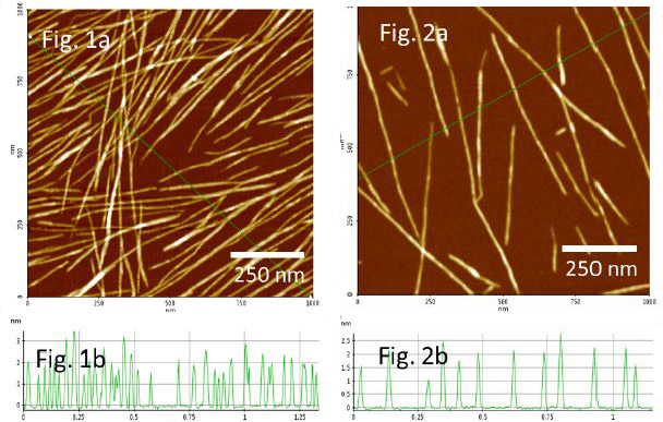
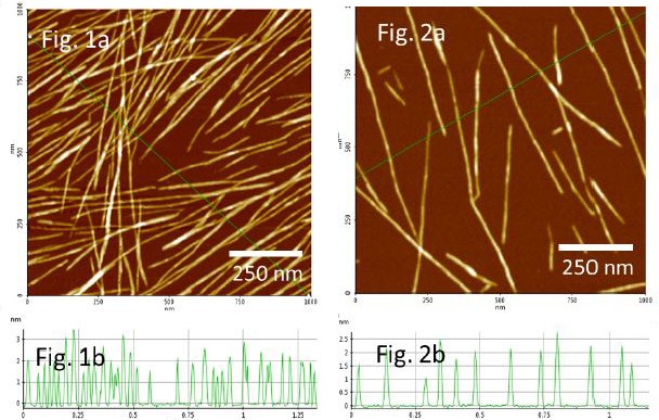
All of the three samples of CNWs showed similar shapes and sizes in AFM topographies. The topography of Sample 1 in Fig. 1a shows the CNW rods are relatively long and straight. The CNWs are approximately 100 to 1000 nm long and 1 to 3 nm wide, as measured from the AFM height profile in Fig. 1b. It is assumed that the CNW rods have circular cross sections and the width is determined by measuring the height of the CNWs. It is interesting to note that the CNWs are well distributed and appear to be oriented along diagonal direction. The air puff that was applied to drive off the residual droplet from the Mica surface was aimed such that the droplet was forced to flow from one side to the opposite side. This rapid droplet flow may have induced the force gradient suited for the CNWs to flow and orient in the direction of the liquid flow.
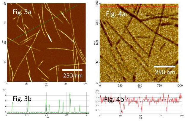
Although not as preferentially oriented, as shown in Figs 2a and 3a, the CNWs of similar shapes can be observed from the AFM topographies of the other two samples, Sample 2 and Sample 3, respectively. They also appear relatively straight and well dispersed but not as densely distributed–bending or entanglement of CNWs were minimal. The width of the CNWs from the Sample 2 and 3 ranging from 1 to 3 nm can be seen from the AFM height profiles of Fig. 2b and 3b, respectively. It is noted that more nanofibrils are seen from the Sample 1 topography than those of the Sample 2 and Sample 3 because the sample 1 was not diluted with DI water as highly as the other two samples.


Image in Fig. 4a is topography of CNWs dispersed on Mica which is overlaid with a color scale of Young’s modulus values measured in the PinPoint mode. In this mode, the forcedistance (f-d) curve is acquired from each pixel in the areas where topography is imaged, and from each f-d curve, elastic modulus is calculated and mapped out in real-time in unison with corresponding topography image. The darker color scale in Fig 4a refers to a lower modulus value. The CNWs are seen darker than surrounding Mica which indicates that the CNWs are not as stiff as the surface of mica. The modulus line profile in Fig 4b shows the modulus value for CNWs is ~180 GPa while that for Mica is ~ 210 GPa. The measured value of the CNF’s Young’s modulus is not too far off of the value of 150 GPa that is predicted theoretically for crystalline cellulose nanowhiskers [7].
Summary
The CNWs were successfully imaged in high resolution and high quality using commercially available AFM. The CNW samples that were well dispersed on Mica without much entanglement allowed for easy measurements of the width, length as well as Young’s modulus of individual CNWs. The results demonstrate that AFM can be used for dimensional nanometrology as well as quantitative nanomechanical property measurements for CNW characterization.
Acknowledgments
Thank you to Dr. Javier Carvajal Barriga from the Pontificia Universidad Católica del Ecuador for the CNW samples for this study and fruitful discussion.
REFERENCES
[1] Yanxia Zhang, Tiina Nypelo, Carlos Salas, Julio Arboleda, Ingrid C. Hoeger, Orlando J. Rojas, J. Renew. Mater., Vol. 1, No. 3, July 2013.
[2] Xuezhu Xu,Haoran Wang,Long Jiang, Xinnan Wang, Scott A. Payne, J. Y. Zhu, and Ruipeng Li⊥, Macromolecules 2014, 47, 3409−3416
[3] Michael T Postek, Andr´as Vlad´ar1, John Dagata1, Natalia Farkas, Bin Ming1, Ryan Wagner, Arvind Raman, Robert J Moon, Ronald Sabo, Theodore H Wegner and James Beecher, Meas. Sci. Technol. 22 (2011) 024005.
[4] Ingrid Hoeger,a Orlando J. Rojas, Kirill Efimenko,c Orlin D. Velevc and Steve S. Kelleya, Soft Matter, 2011, 7, 1957.
[5] Park NX20 - Overview | Park Atomic Force Microscope
http://www.parkafm.com/index.php/products/research-afm/park-nx20/overview
[6] Park AFM Modes
http://www.parkafm.com/index.php/park-afmmodes/nanomechanical-modes
[7] Xiaodong Cao, Youssef Habibi1, Washington Luiz Esteves Magalhães, Orlando J. Rojas, and Lucian A. Lucia, CURRENT SCIENCE, VOL. 100, NO. 8, 25 APRIL 2011 (and references within).
