Redesigning Microwave Impedance Microscopy: Toward Enhanced Nanoscale Dielectric Imaging
- 19 Aug 2025
- Volume 28
- NANOscientific Magazine, 2025
Prof. Eric Y. Ma, Physics, Electrical Engineering & Computer Sciences, University of California, Berkeley
Adapted from Presentation, Edited by NanoScientific
In the realm of scanning probe microscopy, a quiet revolution is taking place—one that aims to democratize high-frequency electrodynamic imaging by radically simplifying the instrumentation without sacrificing sensitivity or spatial resolution. At the 2024 NanoScientific Symposium Americas, Prof. Eric Y. Ma of UC Berkeley introduced a groundbreaking redesign of Microwave Impedance Microscopy (MIM), a technique for mapping local dielectric and conductive properties at the nanoscale. His team’s new architecture eliminates the need for a cancellation circuit and specialized probes—both long-standing technical bottlenecks. Instead, it harnesses self-referenced homodyne detection and commercially available monolithic silicon cantilever probes to achieve thermal-noise-limited sub-zeptofarad sensitivity and electrical spatial resolution limited only by the tip radius. This leap in design not only significantly reduces the cost and complexity barriers to MIM but also unlocks new measurement modalities, such as continuous-frequency local microwave spectroscopy, broadband photoconductivity nano-interferometry, and nonlinear MIM operation.
The Problem with Traditional MIM
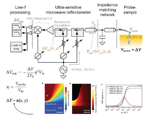
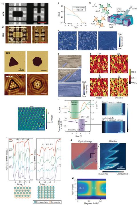
Microwave Impedance Microscopy combines the spatial precision of atomic force microscopy with the electrical sensitivity of microwave reflectometry. It functions by delivering a high-frequency signal (typically in the GHz range) to a nanoscopic probe tip that interacts with the sample via near-field coupling. The reflected signal encodes changes in the tip-sample admittance—dependent on local dielectric constant, conductivity, and depletion/accumulation layers, among others—allowing high-resolution maps of local electronic behavior with minimal sample preparation even for buried samples (Fig. 1). With this unique capability, MIM has made significant contributions to wide-ranging scientific and technology fields (Fig. 2).
But conventional MIM systems face several technical hurdles:
1. Specialized Probes: Traditional MIM relies on highly-specialized, often-handmade, probes designed to minimize parasitic capacitance and signal loss at GHz frequencies. These probes are expensive, fragile, and suffer from relatively large and varying tip radii, severely limiting spatial resolution and robust, quantitative interpretation of MIM images.
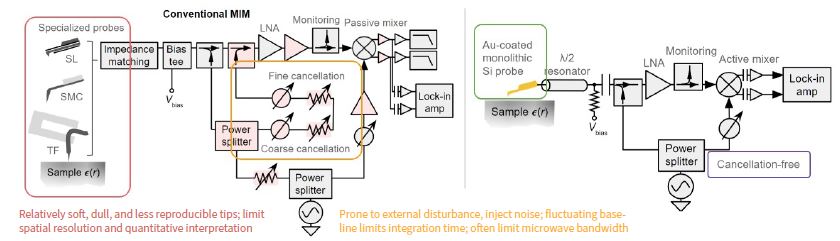
2. Cancellation Circuitry: Another challenge lies in handling the strong reflected baseline signal. Since the signal of interest is a small fluctuation riding on top of a much larger baseline microwave reflection, a cancellation circuit is thought to be necessary to subtract the baseline via destructive interference and avoid amplifier saturation. This cancellation path, however, requires multiple stages of amplitude and phase controllers and is highly susceptible to environmental noise, temperature fluctuations, and vibration.
3. Stability and Complexity: These issues make MIM systems hard to operate stably for long periods to achieve higher sensitivity. Even in controlled lab environments, cancellation often degrades within an hour or two, capping integration time and limiting sensitivity. Moreover, the complexity of both the specialized probes and delicate cancellation circuits makes it challenging to deploy MIM in broader scientific or industrial settings.
A Simpler, More Robust Solution
Prof. Ma’s group proposed a radical simplification: eliminate the cancellation circuit entirely and use standard, metal-coated monolithic silicon AFM cantilever probes (Fig. 3).
• Johnson-noise-limited noise floor, drift-free scan, sub-zF sensitivity, enhanced resolution.
• Makes MIM vastly more accessible
• Facilitates advanced modalities & extensions
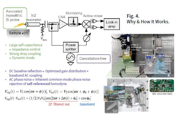
At first glance, this approach seemed counterintuitive. Standard AFM probes have higher self-capacitance and lack shielding, traits traditionally viewed as disqualifying for GHz signal delivery. Without cancellation, the large baseline reflection would saturate the amplification chain or preclude the necessary high signal gain. Yet, by careful impedance design and leveraging the inherent advantages of self-referenced detection and modern AFM probes, the team not only made it work—they achieved new state-of-the-art performance (Fig. 4).
Key Innovations
• Impedance Engineering: The probe structure is integrated into a 50-ohm transmission line by controlling the thickness and dielectric properties of the insulating spacer between the probe carrier chip and the ground plane. This minimizes stray capacitance and maximizes responsivity.
• Dynamic Mode Signal Extraction: Rather than measuring at DC or low frequencies, the MIM signal is extracted at the cantilever’s mechanical resonance frequency (typically hundreds of kilohertz), where noise is reduced.
• AC-Coupled Signal Path: After the reflected signal is down-converted through a self-referenced homodyne mixer, it is AC-coupled, removing the DC baseline without any need for cancellation.
• Common-Mode Noise Rejection: The self-referenced homodyne detection scheme ensures that shared phase noise between the reference and signal paths cancels out, greatly reducing noise in the baseband output.
• Tighter Integration: All RF signal conditioning, demodulation, and baseband amplification electronics, including baluns, an active mixer and low-noise instrumentation amplifiers, are integrated onto a single printed circuit board. This minimizes wiring losses, thermal drift, and noise pickup.
Benchmarking Against Commercial Systems
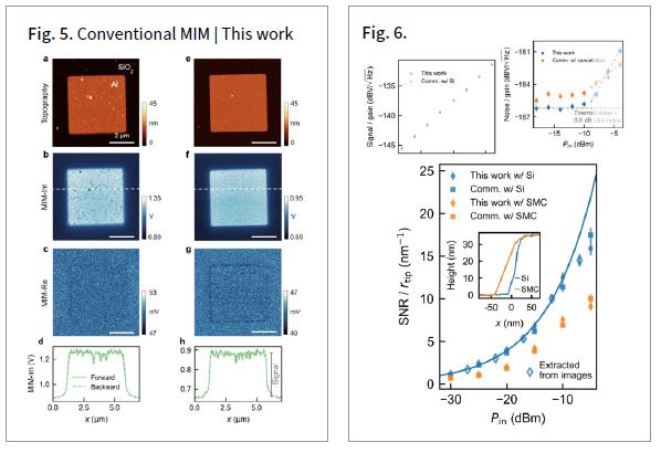
To assess performance, the team performed side-by-side experiments using both their cancellation-free system with monolithic silicon probes and an expensive, cutting-edge commercial MIM platform. The test sample was an array of thin aluminum dots deposited on a silicon dioxide substrate—chosen for its strong capacitive contrast and sharp material boundaries (Fig. 5).
Results:
• Improved Spatial Resolution: Thanks to the smaller (and more consistent) tip radius of commercial silicon AFM cantilevers, the new system resolved features more crisply, particularly small dust particles and edges, in both topography and MIM.
• Higher MIM signal-to-noise-ratio (SNR): The SNR in the MIM channels are comparable, if not better, despite a sharper tip and the absence of cancellation or shielding.
Breaking the Sensitivity Barrier: Thermal Noise-Limited Operation and Long Averaging Time
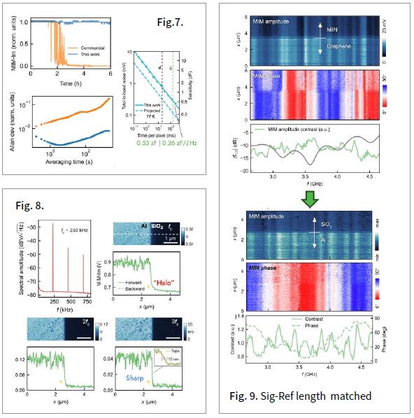
One of the most striking findings from Prof. Ma’s group was that their system operated at the Johnson-Nyquist thermal noise limit for powers below approximately −10 dBm. This means the system’s sensitivity is constrained only by fundamental physics, not by technical or environmental noise(Fig. 6).
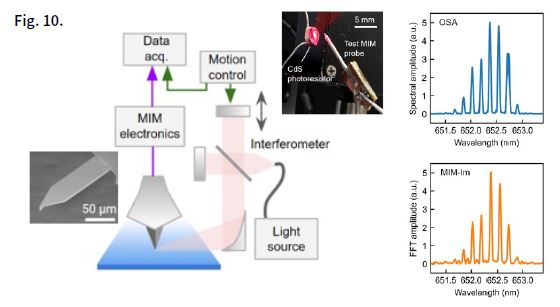
The implications are profound. It suggests that the system has reached the theoretical maximum sensitivity for room temperature operation—and that any further improvement in noise performance must come from cooling the combined tip-sample system and the microwave preamplifiers. Moreover, the cancellation-free architecture eliminates drift-induced saturation completely and enables significantly longer data acquisition times, resulting in higher SNR and improved sensitivity. In a 6.5-hour real-world scan of a graphene monolayer encapsulated in hexagonal boron nitride (hBN), the system detected minute variations in subsurface conductivity with an absolute capacitive sensitivity of 0.53 zF (1 zF = 10-21 farad), corresponding to a normalized sensitivity of 0.26 zF/√Hz—a new record for capacitance-based scanning probe technique, to the best of the group’s knowledge (Fig. 7).
Enhanced Spatial Resolution via Harmonic Demodulation
Beyond improving sensitivity, the much higher baseband bandwidth (~1 MHz) of the new architecture also supports higher spatial resolution through harmonic demodulation. By extracting MIM signals not only at the cantilever’s fundamental mechanical resonance but also its 2nd and 3rd harmonics, the team revealed increasingly localized electrical interactions.
This enabled them to resolve sharper transitions in conductivity with spatial resolution comparable to the topographic limit imposed by the tip radius, while suppressing artifacts such as halo effects seen in traditional MIM images.
New Modalities: Expanding What MIM Can Measure
1. Continuous-Frequency MIM Spectroscopy
Conventional MIM spectroscopy typically requires changing hardware and re-tuning multiple amplitude and phase controllers in the cancellation line to switch frequencies. This makes multi-frequency measurements cumbersome and inconsistent. With the new architecture, Prof. Ma’s group demonstrated continuous-frequency spectroscopy using a broadband resistive matching network coupled with matching signal and reference path lengths. In early data, they measured spatially resolved amplitude and phase variations between 2.7 and 4.6 GHz at a metal-insulator boundary—paving the way for more detailed modeling of frequency-dependent permittivity in nanostructures (Fig. 9).
2. Broadband Photoconductivity with Fourier Transform MIM
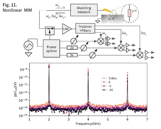
In a novel adaptation, the group used the MIM tip as a localized photoconductivity detector in a Fourier-transform interferometer. By focusing modulated light onto the tip-sample junction and capturing the resulting interferogram in the MIM signal, they extracted spectrally resolved photoresponse data with nanoscale spatial resolution and picometer-scale spectral resolution (Fig. 10).
3. Nonlinear MIM via Harmonic Detection
Finally, the team developed a formalism to interpret MIM signals at 2nd and 3rd harmonics of the excitation frequency, using concepts from nonlinear optics. By solving nonlinear admittance equations, they linked these harmonics to higher-order dielectric and conductive responses of materials—offering a powerful means of probing the rich information contained in local electrical nonlinearities, particularly relevant to semiconductors, strongly-correlated materials, and superconductors.
Implications and Outlook
The redesigned MIM architecture represents a transformative shift in non-invasive nanoscale electronic imaging. It dramatically reduces cost and complexity barriers while improving performance in both sensitivity and spatial resolution—making MIM accessible to broader research and industrial communities.
Future directions include integration with techniques such as STM, NSOM, and MFM, as well as operation at cryogenic temperatures to achieve yoctofarad (10⁻²⁴ F) sensitivity. Additional goals involve applying MIM in biological and aqueous environments, developing highly quantitative methods for extracting local conductivity or doping concentrations, and enabling broader commercial deployment.
As Prof. Ma concluded, “By simplifying the system and letting the physics shine through, we’ve opened the door to broader adoption, deeper and more quantitative insights, and entirely new kinds of measurements that were previously out of reach.”

Dr. Ma is an Assistant Professor in Physics and EECS and holds the Georgia Lee Chair in Physics. He is also a Faculty Scientist at the Lawrence Berkeley National Laboratory. His research focuses on
probing and controlling wave-matter interactions in condensed matter systems at deeply sub-wavelength scales using advanced instrumentation and AI/ML techniques. As an educator, Eric is passionate about using modern software and hardware tools to
enhance undergraduate physics research and education,
especially in laboratory courses.
For more information visit https://sites.google.com/berkeley.edu/ma-lab
References
1. M. E. Barber, E. Y. Ma, and Z.-X. Shen, Nat. Rev. Phys. 4, 61 (2022).
2. J.-Y. Shan, A. Pierce, and E. Y. Ma, Appl. Phys. Lett. 121, 121601 (2022).
3. J.-Y. Shan, N. Morrison, and E. Y. Ma, Appl. Phys. Lett. 122, 121601 (2023).
4. J.-Y. Shan, N. Morrison, S.-D. Chen, F. Wang, and E. Y. Ma, Nat. Commun. 15, 5043 (2024).
5. J. Shan, N. Morrison, and E. Y. Ma, in 2024 IEEE/MTT-S International Microwave Symposium (IMS) (IEEE, 2024), pp. 994–997.
6. A. Y. Waghmare, J. Bromley, J.-Y. Shan, and E. Y. Ma, Appl. Phys. Lett. 126, 131601 (2025).
7. Z. Chu, L. Zheng, and K. Lai, Annu. Rev. Mater. Res. 50, 105 (2020).
