Pushing The Limits: Deeper Insights In PFM and a New Dimension In MFM
- 03 Nov 2025
- Volume 29
- NANOscientific Magazine, 2025
Prof. Dr. Lukas M. Eng, Institute of Applied Physics, TU Dresden,
Germany Adapted from Presentation, Edited by NanoScientific
Introduction
At the 2024 NanoScientific Forum Europe in Munich, Prof. Dr. Lukas M. Eng brought the audience back to two of the most established techniques in scanning probe microscopy—Piezoresponse Force Microscopy (PFM) and Magnetic Force Microscopy (MFM)—only to show how both are being pushed far beyond their traditional limits. For more than thirty years, these techniques have been the workhorses for imaging ferroelectric and magnetic domains at the nanoscale. But Prof. Eng’s latest work at TU Dresden demonstrates how careful experimental design and theoretical modeling can expand what these methods can do. In PFM, his group has been asking a deceptively simple question: how far into a material can PFM “see” beneath the surface? And in MFM, they are extending the method from its classic out-of-plane sensitivity into a true three-dimensional probe of magnetic fields. The results redefine the boundaries of both techniques—turning PFM into a quantitative volumetric probe and MFM into a multidimensional magnetic field mapping tool.
PFM: Looking Beneath the Surface
PFM has long been a go-to technique for visualizing ferroelectric domain structures. Since its origins in the early 1990s, when K. Franke first demonstrated voltage-modulated scanning force microscopy [1], the method has been refined and expanded. Prof. Eng himself played a key role in this evolution, introducing in-plane PFM in 1998 [2] and full three-dimensional PFM the following year [3]. Yet despite decades of use, one fundamental question persisted: when we apply an AC bias to the tip in PFM, what is the actual volume of the material we are probing? Most PFM measurements are interpreted as surface-sensitive, but there have been hints for years that the electric field penetrates far into the bulk. Quantifying that penetration depth has been a long-standing challenge. Earlier efforts provided glimpses of the answer. In 2009, Johann and co-workers showed that in lithium niobate, measurable PFM signals could be detected to depths of roughly 1.7 microns [4]. In 2019, Steffes et al. introduced tomographic PFM [5], physically milling away the sample layer by layer to correlate PFM signals with depth. While effective, the method was destructive and impractical for most applications. Prof. Eng’s group sought a different path [6]: a way to measure the effective probing depth (see Fig. 1) of PFM quantitatively, without damaging the sample.
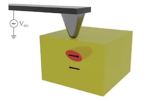
into the depth of a sample.
The work was led by Ph.D. candidate Matthias Roeper, who developed a theoretical model describing how the AC electric field applied by the PFM tip penetrates into a dielectric. In an idealized scenario, with no free charges, the field could extend indefinitely into the bulk. In reality, geometry matters. The model showed that solely the tip radius and the signal-to-noise performance of the instrument defines the effective probing depth and depth resolution. The model as shown in Fig. 2 predicts that the PFM signal is at its maximum close to the sample surface, then reaches a zero-amplitude value at a depth equal to the tip radius, before recovering into the sample depth. The 90-percent depth—defined as 90-percent of the initial normalized PFM amplitude signal at the sample surface—is achieved at nineteen times the tip radius. This 90-percent probing depth can be used to estimate the effective probing depth for standard PFM cases in a much simpler way, rather than applying the full calculation using a multi-parameter model.
Testing the model experimentally required a carefully prepared sample. The group chose x-cut periodically-poled lithium niobate (PPLN), a crystal whose domains are polarized in-plane (see Fig. 2). By embedding the crystal in epoxy and polishing it at a shallow wedge of about three degrees, they created a surface where domain boundaries appeared at progressively greater depths along the scan direction. This allowed the PFM tip to encounter increasing depths in a single scan.
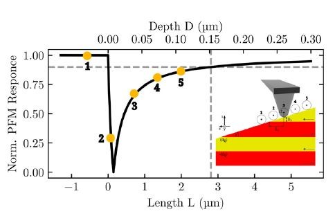
The results aligned strikingly with the model. The PFM amplitude showed a sharp change when the tip reached a depth equal to its radius, and then recovered hyperbolically as the depth increased. The 90-percent probing depth matched the predicted nineteen times of the tip radius. For a typical 60-nanometer platinum-coated tip, the method could non-destructively probe more than one micron into the sample. One of the strengths of the model was its robustness. The team systematically varied the AC voltage from one to above eight volts, changed the drive frequency between resonance and off-resonance conditions, and altered the contact force from light to heavy. None of these changes affected the measured depth. Only the tip radius mattered—a simple but powerful insight.
Prof. Eng emphasized one important caveat for researchers working on thin films and two-dimensional materials. In ultrathin samples, the PFM field penetrates well beyond the active layer into the substrate. The measured signal will therefore include contributions from the substrate, making it essential to account for substrate properties when interpreting PFM data from van der Waals ferroelectrics or oxide heterostructures.
MFM: Adding a New Dimension
While the advance in PFM focuses on looking deeper into a material, the innovation in MFM aims to look in more directions at once.
Conventional MFM is typically performed in vertical mode (i.e., VMFM), in which the cantilever oscillates in its flexural mode to detect the out-of-plane component of the magnetic stray field above a sample (see Fig. 3a). This works well for simple ferromagnetic domain structures, such as those in a hard disk, but it captures only part of the picture. In modern magnetic materials—particularly those hosting topological spin textures such as skyrmions and antiskyrmions—the in-plane components of the stray field are equally important for understanding the underlying physics.
Prof. Eng’s group set out to extend MFM into three dimensions by measuring both the vertical and lateral components of the magnetic field (see Fig. 3a). This work, led by Bachelor’s student Jori Schmidt and supervised by postdoc Sam Seddon, involved exciting the cantilever in both its flexural and torsional resonances simultaneously [7]. The flexural resonance captures the vertical component (MFM or VMFM) while the torsional resonance captures the in-plane components (LMFM). With separate piezo excitations and lock-in amplifiers for each mode, both signals can be collected at once.
Testing the approach on a hard disk with well-defined perpendicular magnetic domains provided a clear proof of concept (Fig. 3). In VMFM mode, the alternating up and down magnetization of the domains produced the expected stripe contrast. In LMFM mode, strong contrast appeared only when the torsional oscillation was perpendicular to the domain walls (see Fig. 3b,c), whereas the contrast disappeared when the oscillation was aligned parallel to the walls. This matched the expected magnetic symmetry and confirmed that LMFM was selectively detecting the appropriate in-plane field components.
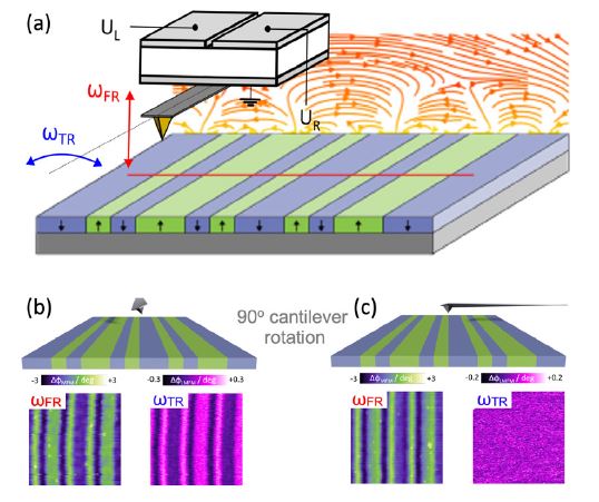
To validate the interpretation, the group modeled the MFM tip as a magnetic point dipole using the magpylib simulation package. The simulated stray fields produced VMFM and LMFM contrasts in close agreement with the experimental measurements, including the phase inversion between the vertical and lateral signals. Small discrepancies for LMFM when the torsional oscillation was parallel to the domain walls were attributed to subtle tip–sample coupling effects, which will be explored in future refinements.
The significance of 3D MFM becomes clear in more complex materials. Prof. Eng’s group has already started to apply this technique to MnPtPdSn samples, which host antiskyrmion lattices at room temperature. These topological spin textures cannot be fully understood without measuring both vertical and in-plane components of the stray field. By thinning lamellae to around two microns and mounting them free-standing, the group could perform 3D MFM to capture the complete stray field distribution.
Conclusion: Extending the Reach of Scanning Probe Microscopy
In PFM, Prof. Eng’s group has transformed the question of depth from a vague concept into a quantitative, non-destructive measurement directly linked to the tip radius and the instrument’s performance. In MFM, they have expanded the technique into a true three-dimensional probe capable of revealing complex magnetic field distributions. These developments will have broad implications for researchers working in (multi-)ferroic materials, where functionality often depends on structures buried below the surface or on the interplay of multiple field components. Prof. Eng’s work serves as a reminder that even well-established techniques can be extended to new frontiers when carefully examined.
As he told the NanoScientific Forum Europe audience, “In PFM, remember the substrate. In MFM, remember the other components of the field.” It is a fitting summary of an approach that balances experimental precision with physical insight—pushing the limits of scanning probe microscopy into deeper and more multidimensional territory.
About Prof. Dr. Lukas Eng
Dr. Lukas M. Eng is a Full Professor at the Excellence University “Technische Universität Dresden (TUD), Germany.” His research focuses on experimentally exploring the nanoscale magnetic, electronic, and optical properties of modern low-dimensional and/or topological materials by applying various scanning-probe-microscopy-based methods at both room and low temperatures. A particular emphasis is placed on investigating frustrated nanomagnetic systems that exhibit non-collinear spin textures such as skyrmions, as well as photon-polaritons in layered and twisted 2D materials.
He earned his Ph.D. and venia legendi from the University of Basel, Switzerland, before being appointed as a full professor at TUD following several postdoctoral positions—namely in Molecular Electronic Devices (BASF AG, Germany), Biophysics (University of Geneva, Switzerland), and Nonlinear and Quantum Optics (ETH Zürich, Switzerland).
Prof. Eng has published over 400 peer-reviewed papers in fields including nano-optics, nanoelectronics, non-collinear magnetic nanostructures, linear and nonlinear optics, and biophysics.
More information is available at: https://tudresden.de/mn/physik/iap/experimentalphysik-photophysik#intro.
References
-
K. Franke, L. M. Eng, M. Weihnacht, W. Haessler, and J. Besold. The creation of the piezoresponse force microscopy twenty-three years ago. IEEE Intern. Symp. Appl. Ferroelectr. & Europ. Conf. Appl. Polar Dielectr. and Piezoelectric Force Microscopy Workshop (ISAF/ECAPD/PFM), 1 (2016); https://doi.org/10.1109/ISAF.2016.7578100.
-
M. Abplanalp, L. Eng, and P. Günter. Mapping the Domain Distribution at Ferroelectric Surfaces by Scanning Force Microscopy. Appl. Phys. A 66, S231 (1998); https://doi.org/10.1007/s003390051136.
-
L. M. Eng, H.-J. Güntherodt, G. A. Schneider, U. Köpke, and J. Muñoz Saldaña. Nanoscale Reconstruction of Surface Crystallography from 3-Dimensional Polarization Distribution in Ferroelectric Barium-titanate Ceramics. Appl. Phys. Lett. 74, 233 (1999); https://doi.org/10.1063/1.123266.
-
F. Johann, Y. J. Ying, T. Jungk, Á. Hoffmann, C. L. Sones, R. W. Eason, S. Mailis, and E. Soergel. Depth resolution of piezoresponse force microscopy. Appl. Phys. Lett. 94, 172904 (2009).
-
J. J. Steffes, R. A. Ristau, R. Ramesh, and B. D. Huey. Thickness scaling of ferroelectricity in BiFeO₃ by tomographic atomic force microscopy. Proc. Natl. Acad. Sci. U.S.A. 116, 2413–2418 (2019).
-
M. Roeper, S. D. Seddon, Z. H. Amber, M. Rüsing, and L. M. Eng. Depth resolution in piezoresponse force microscopy. J. Appl. Phys. 135, 224102 (2024); https://doi.org/10.1063/5.0206784.
-
J. Schmidt, L. M. Eng, and S. D. Seddon. Towards 3D Magnetic Force Microscopy: Simultaneous torsional cantilever excitation to access a second, orthogonal stray field component. J. Appl. Phys. 136, 113904 (2024); https://doi.org/10.1063/5.0226570.
Category