Imaging Forbidden Light: Nano-Spectroscopy Of Polaritons In 2d Oxide Membranes
- 10 Nov 2025
- Volume 29
- NANOscientific Magazine, 2025
Prof. Alex McLeod, University of Minnesota
Adapted from Presentation, Edited by NanoScientific
Introduction: Probing the Hidden Realm of Light and Matter
At the 2024 NanoScientific Symposium Americas, Prof. Alex McLeod of the University of Minnesota invited attendees into a very different vision of optical microscopy—one that ventures deep into a regime traditional optics cannot reach. His focus was on polaritons—hybrid light–matter excitations that live in the so-called “forbidden light” region—and on the technique capable of imaging them: scattering-type scanning near-field optical microscopy (s-SNOM). In this “zone of forbidden light,” near-field optical techniques make visible what far-field optics cannot. Polaritons not only provide a window into the quantum properties of materials, but can also be engineered and manipulated for advanced photonic applications. Prof. McLeod’s research reveals how these exotic excitations propagate, interact, and even refract in man-made 2D oxide membranes (Fig. 1).
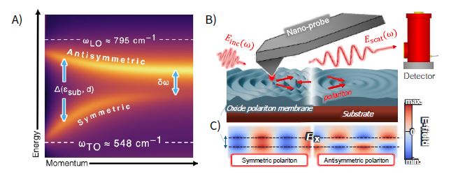
Fig. 1.A) Phonon polaritons in SrTiO₃ membranes are hybrid modes of light and lattice vibrations.Coupling across the membrane’s top and bottom interfaces forms symmetric and antisymmetric modes with tunable positive or negative dispersion of energy (ω) versus momentum (q ∝ λ, where λ is the polariton wavelength). B) Nearfield microscopy uses a sharp, metal-coated probe illuminated by an infrared laser to launch and detect polaritons by scattering their evanescent fields to a detector.Veselago lensing occurs when symmetric polaritons enter a region with negative dispersion.C) The internal distribution of oscillating charges and lateral electric fields (e.g., Ex) determines the mode symmetry.
Quantum Materials: Why Resolution Matters
Prof. McLeod’s scientific motivation begins with a broad category: quantum materials—systems in which interactions between electrons, or between electrons and the crystal lattice, dominate their physical behavior. This interaction-driven complexity produces emergent phenomena such as insulator–metal transitions in oxides like VO₂, driven by temperature or external fields; high-temperature superconductivity, often competing with charge-ordered phases; topological edge states with spin-selective conduction; and designer quantum phases, such as twisted bilayer graphene that hosts superconductivity at “magic” angles.
However, these phenomena are spatially heterogeneous. Domains form, percolate, and compete at nanometer scales. Averages over microns or millimeters miss these fine details.
This is where high-resolution scanning probe techniques—AFM, STM, and in Prof. McLeod’s case, near-field optical spectroscopy—become essential. They not only resolve local properties but can optically map phase coexistence and transitions in real space.
Beating the Abbe Limit: Why Forbidden Light Matters
For optical scientists, the Abbe diffraction limit has long been a barrier: the smallest resolvable feature in conventional optics is proportional to the wavelength of light. For the mid-infrared light Prof. McLeod uses to probe low-energy excitations, that limit is on the order of microns—far too coarse for nanoscale physics.
The solution is to enter the near field. A sharp, metalized AFM tip is illuminated with infrared light, acting as a nanoscopic antenna that localizes the field at its apex. The sample’s response is encoded in evanescent waves—non-propagating fields confined to nanometer distances from the surface. These near-field signals are scattered by the tip into the far field, where they can be collected by a detector.
This approach lets Prof. McLeod image “forbidden light”—evanescent modes, including polaritons, that cannot be accessed with standard far-field optics.
Polaritons: Hybrid Waves of Light and Matter
The stars of Prof. McLeod’s work are polaritons—mixed modes that combine electromagnetic waves with material excitations. Depending on the host material, these can include:
• Plasmon polaritons (light coupled to free electron oscillations)
• Phonon polaritons (light coupled to lattice vibrations)
• Exciton polaritons (light coupled to bound electron–hole pairs)
• Magnon polaritons (light coupled to spin waves)
In all cases, polaritons compress the wavelength of light, sometimes by factors of 100 or more, allowing them to propagate and interact at scales ideally suited for nanophotonics.
Because they live in the near field, polaritons are highly confined and long-lived—immune to direct radiation loss into free space unless scattered by edges or defects. This makes them excellent candidates for on-chip guiding, focusing, and sensing.
Polariton Interferometry: Measuring Wavelength and Lifetime
To characterize polaritons, Prof. McLeod’s team uses interferometric mapping. The AFM tip launches polaritons into the material, where they propagate until reflecting from an edge or interface. The forward and reflected waves interfere, producing a standing wave pattern.
By scanning the tip toward and away from an edge, Prof. McLeod records interference fringes. From these fringes, the polariton wavelength and propagation length (related to the quality factor Q) can be extracted.
In high-quality graphene encapsulated in hexagonal boron nitride, for example, Q-factors can exceed 50–100 at low temperature. In oxide membranes, current Q-factors are lower (~3) due to scattering, but the technique remains the most direct way to study polariton behavior.
Moving Beyond Graphene: Why Oxide Membranes?
Graphene has been a showcase for plasmon polaritons, but Prof. McLeod’s current interest is in transition metal oxides, particularly strontium titanate (SrTiO₃). Like other so-called perovskite oxide crystals, SrTiO₃ showcases ferroelectricity—or “switchable” macroscopic electric polarization—but also more exotic behaviors like unconventional superconductivity, metal–insulator transitions, and strong coupling between lattice, charge, and orbital degrees of freedom.
These rich interactions make oxides an ideal platform to explore how light–matter coupling can be tuned, enhanced, or even reversed. By fabricating freestanding oxide membranes only tens of nanometers thick, Prof. McLeod’s team can precisely control boundary conditions, enabling new hybrid modes and optical responses that are impossible in bulk crystals.
Moving Beyond Graphene: Why Oxide Membranes?
Graphene has been a showcase for plasmon polaritons, but Prof. McLeod’s current interest is in transition metal oxides, particularly strontium titanate (SrTiO₃). Like other so-called perovskite oxide crystals, SrTiO₃ showcases ferroelectricity—or “switchable” macroscopic electric polarization—but also more exotic behaviors like unconventional superconductivity at low temperature.
These oxides are grown by molecular beam epitaxy (MBE) onto sacrificial buffer layers. Using remote epitaxy, the oxide film can then be peeled from the growth substrate and transferred as a free-standing membrane onto almost any target substrate. Prof. Bharat Jalan at the University of Minnesota, Prof. McLeod’s chief collaborator, has pioneered hybrid MBE techniques to grow a wide assortment of oxides in membrane form. This opens new frontiers for nanoelectronics and nanophotonics based entirely on quasi-2D oxides.
These materials offer several advantages, including custom thicknesses ranging from a few to tens of nanometers, bulk-like crystallinity even at small thicknesses, and the ability to tailor substrate environments to control polariton dispersion. They also exhibit switchable phases such as ferroelectricity, which are not easily achieved in conventional 2D materials. Additionally, oxides like SrTiO₃ host strong infrared-active phonons, making them ideal for phonon polariton generation and manipulation.
Hyperspectral Nano-Imaging: Mapping Polaritons in Action
Using a Park Systems NX10 AFM coupled with a tunable pulsed laser (via optical parametric amplification), Prof. McLeod performs hyperspectral nano-FTIR imaging.
This system combines:
• Broadband frequency coverage for phonon resonances
• Michelson interferometer for phase-resolved detection
• Tapping mode operation for simultaneous AFM and optical mapping
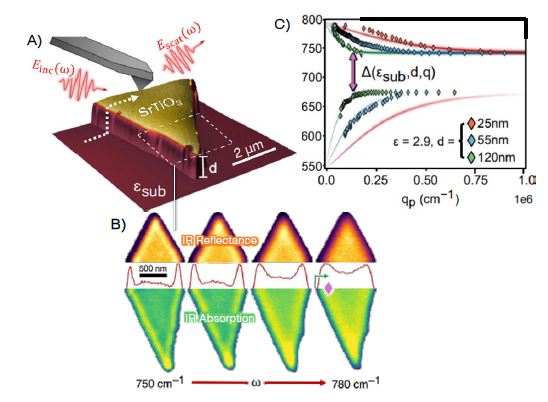
In SrTiO₃ membranes, the team images phonon polariton fringes emanating from edges. By tuning the laser frequency, they record the dispersion relation—how the polariton wavelength changes with frequency (Fig. 1A).
This measurement directly confirms the presence of propagating phonon polaritons (Fig. 2A–B) and matches theoretical models based on the membrane thickness and dielectric environment.
Two-Branch Polariton System: Symmetric and Antisymmetric Modes
A key discovery is that oxide membranes support two polariton modes. Each surface (top and bottom) can host a surface polariton. When the two interact, they form:
• Symmetric (bonding) mode with positive group velocity
• Antisymmetric (antibonding) mode with negative group velocity
Fig. 1C shows the predicted electric field profile of these interfacially coupled hybrid polariton modes. The separation between these modes is a Rabi splitting (Δ) proportional to their mutual coupling strength, measured directly in Fig. 2C.
The opposite group velocities are particularly intriguing. Negative group velocity polaritons open the door to negative refraction, or Veselago lensing—where polariton waves focus beyond a lens without diffraction (Fig. 1B).
Veselago Lensing in Membranes
By engineering suspended and supported regions of the membrane, Prof. McLeod can spatially control polariton dispersion.
In one configuration:
• Suspended region supports propagating modes
• Supported region creates a polaritonic “bandgap”
A polariton launched from a point source in the suspended region can refract negatively at the interface, focusing into a sharp image in the supported region.
This on-chip Veselago lens could form the basis for polariton optics devices, enabling subwavelength focusing and imaging.
Instrumentation: A Platform for Forbidden Light
The experiments require a carefully integrated system built upon a Park Systems scanned probe microscope:
• Park Systems NX10 AFM for stable tapping-mode near-field operation
• Parabolic mirror + retroreflector for focusing illumination and collecting scattered light
• Michelson interferometer for phase and amplitude separation
• Broadband tunable pulsed laser covering mid-infrared phonon energies
• Cryogenic stage for low-temperature measurements (down to 15 K) to improve Q-factors
The setup allows simultaneous topography, optical maps, and electrical potential mapping—enabling true multi-modal characterization of quantum materials.
Challenges and Future Directions
Current polariton Q-factors in oxide membranes are modest, limited by scattering from imperfections. Improving MBE growth, membrane transfer, and surface quality will help. Cryogenic measurements are expected to extend propagation lengths significantly. Combined with membrane engineering (thickness, substrate tuning), this will open the way to on-chip polariton waveguides, resonators, and logic circuits.
Conclusion: A New Kind of Optics for Quantum Materials
Prof. Alex McLeod’s work marks a shift in optical microscopy—from imaging surfaces to imaging the light trapped within them. By mapping polaritons in 2D oxide membranes, his team is developing a powerful platform for mid-infrared nanophotonics, potentially transforming how we sense, process, and control quantum materials at the nanoscale.
These hybrid light–matter waves not only reveal hidden optical behavior but also open practical pathways toward compact, chip-scale devices for spectroscopy, sensing, and photonic computation.
As he told the NSFA audience: “We’re not just imaging materials anymore. We’re imaging their forbidden light—and in doing so, opening entirely new dimensions of control over quantum matter.”

About Prof. Alex McLeod
Dr. Alex McLeod is an Assistant Professor in the School of Physics and Astronomy at the University of Minnesota, where he leads research in nano-optical spectroscopy and quantum materials. He earned his Ph.D. in Physics with honors from the University of California, San Diego, and completed postdoctoral work at Columbia University as a Director’s Fellow with the Columbia Nano Initiative.
His research explores phase transitions in quantum matter and the optoelectronic properties of next-generation 2D materials, including graphene and van der Waals heterostructures. Dr. McLeod designs and builds advanced near-field optical instruments—often operating at cryogenic temperatures—to probe light–matter interactions at the nanoscale.
By combining precision experiments with theoretical analysis, his work has advanced understanding in low-temperature polaritonics and non-destructive nanospectroscopy, including contributions to NASA-supported sample analysis missions. Prof. McLeod is recipient of the 2022 International Union for Pure and Applied Physics (IUPAP) Young Faculty Award for his studies of polaritons and nanoscale phase transitions at low temperatures.
References
-
Lukaskawcez, B.; Varshney, S.; Choo, S.; Park, S. H.; Seo, D.; Thompson, L.; Hirshberg, N.; Garber, M.; Uram, D.; Binger, H.; Koester, S.; Oh, S.-H.; Low, T.; Jalan, B.; McLeod, A. Interfacial Strong Coupling and Negative Dispersion of Propagating Polaritons in Freestanding Oxide Membranes. arXiv, June 27, 2025.
-
Qazilbash, M. M.; Brehm, M.; Chae, B. G.; Ho, P. C.; Andreev, G. O.; Kim, B. J.; Yun, S. J.; Balatsky, A. V.; Maple, M. B.; Keilmann, F.; Kim, H. T.; Basov, D. N. Mott Transition in VO₂ Revealed by Infrared Spectroscopy and Nano-Imaging. Science 2007, 318 (5857).
-
Chen, X.; Hu, D.; Mescall, R.; You, G.; Basov, D. N.; Dai, Q.; Liu, M. Modern Scattering-Type Scanning Near-Field Optical Microscopy for Advanced Material Research. Advanced Materials 2019, 31 (24).
-
Novotny, L. Optics InfoBase: Journal of the Optical Society of America A – Allowed and Forbidden Light in Near-Field Optics. I. A Single Dipolar Light Source. JOSA A 1997, 14 (1), 91–104.
-
Basov, D. N.; Fogler, M. M.; García De Abajo, F. J. Polaritons in van der Waals Materials. Science 2016, 354 (6309).
-
Fei, Z.; Rodin, A. S.; Andreev, G. O.; Bao, W.; McLeod, A. S.; Wagner, M.; Zhang, L. M.; Zhao, Z.; Thiemens, M.; Dominguez, G.; Fogler, M. M.; Castro Neto, A. H.; Lau, C. N.; Keilmann, F.; Basov, D. N. Gate-Tuning of Graphene Plasmons Revealed by Infrared Nano-Imaging. Nature 2012, 486 (7405).
-
Ni, G. X.; McLeod, A. S.; Sun, Z.; Wang, L.; Xiong, L.; Post, K. W.; Sunku, S. S.; Jiang, B. Y.; Hone, J.; Dean, C. R.; Fogler, M. M.; Basov, D. N. Fundamental Limits to Graphene Plasmonics. Nature 2018, 557 (7706), 530–533.
-
Varshney, S.; Choo, S.; Thompson, L.; Yang, Z.; Shah, J.; Wen, J.; Koester, S. J.; Mkhoyan, K. A.; McLeod, A. S.; Jalan, B. Hybrid Molecular Beam Epitaxy for Single-Crystalline Oxide Membranes with Binary Oxide Sacrificial Layers. ACS Nano 2024, 18 (8), 6348.
-
Steinle, T.; Mörz, F.; Steinmann, A.; Giessen, H. Ultra-Stable High Average Power Femtosecond Laser System Tunable from 1.33 to 20 μm. Optics Letters 2016, 41 (21).
-
Lewin, M.; Baeumer, C.; Gunkel, F.; Schwedt, A.; Gaussmann, F.; Wueppen, J.; Meuffels, P.; Jungbluth, B.; Mayer, J.; Dittmann, R.; Waser, R.; Taubner, T. Nanospectroscopy of Infrared Phonon Resonance Enables Local Quantification of Electronic Properties in Doped SrTiO₃ Ceramics. Advanced Functional Materials 2018, 28 (42), 1802834.
-
Huth, F.; Govyadinov, A.; Amarie, S.; Nuansing, W.; Keilmann, F.; Hillenbrand, R. Nano-FTIR Absorption Spectroscopy of Molecular Fingerprints at 20 nm Spatial Resolution. Nano Letters 2012, 12 (8), 3973–3978.
Category