
Where Nanotechnology Shapes the Future of Science
Explore cutting-edge breakthroughs and connect with the world's leading nanoscience researchers.
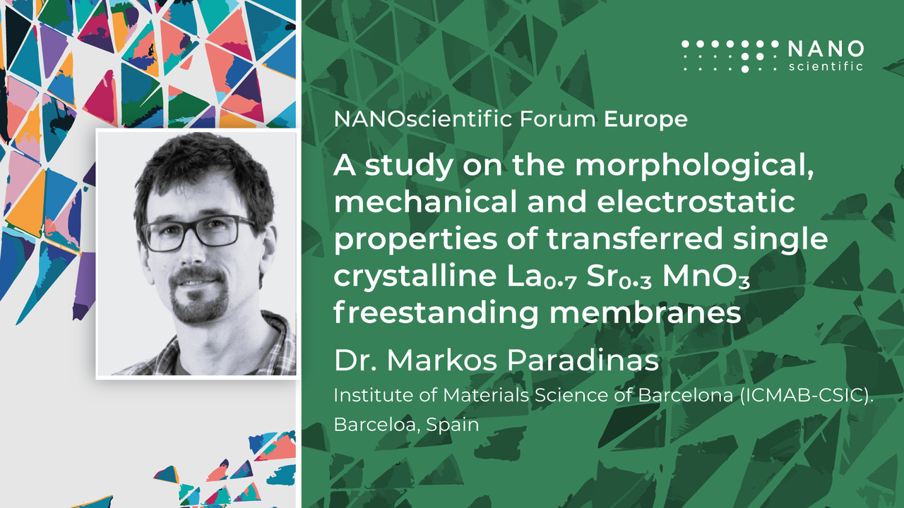
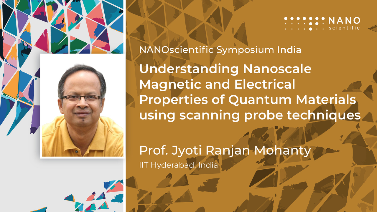
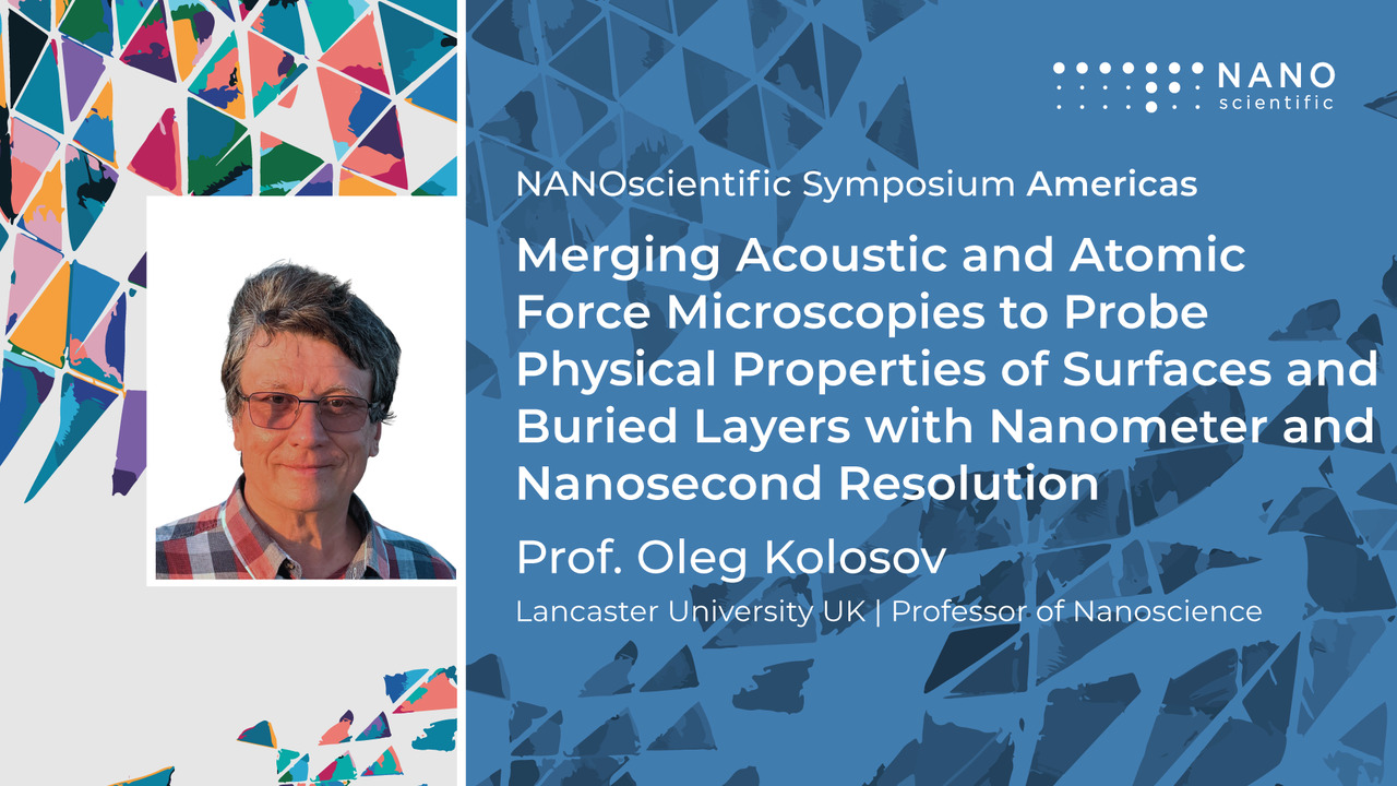
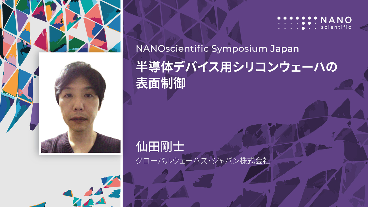
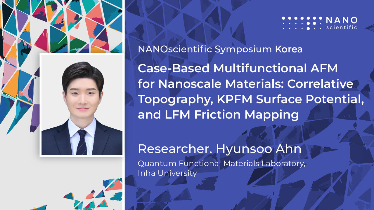
NANOscientific Symposium
Connecting the Global Community of Nanoscience Experts
NANOscientific Symposium brings together experts from around the globe to explore the science, engineering, and applications of nanotechnology. Featuring keynote addresses and virtual networking opportunities, attendees can form connections and propel their research, innovation, and businesses forward.
NANOscientific Magazine
Exploring the Full Spectrum of Nanotechnology
Free for researchers and professionals worldwide — covering the latest in nanoscience, microscopy, and nanotechnology since 2014.
On-Demand
Nanoscience Presented by the World's Leading Researchers
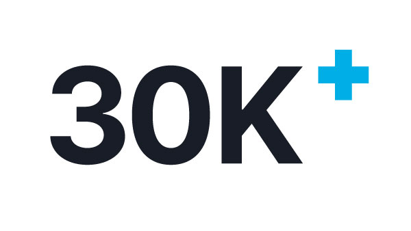
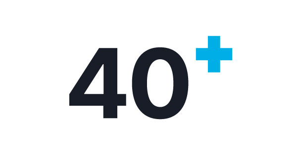
Every NANOscientific Symposium session is archived and freely available to watch. Browse presentations from researchers across 40+ countries, organized by topic and region.
By the Numbers
Trusted by the Global Nanoscience Community
A global platform connecting nanoscience researchers, industry leaders, and innovators through annual symposiums and publication.


Programs & Resources
Everything You Need to Advance Your Research
From recorded symposium sessions to online AFM courses and scholarship opportunities, NANOscientific provides the resources researchers need at every stage.